Rhizosphere Influence on Plant Medicine

Einjähriger Beifuß (Artemisia annua)
Artemisia annua
Wikipedia

Mycorrhization leads to nutrient and information flow, often in both directions. The plant root supplies sugars to the fungus, while the fungus induces Jasmonic Acid biosynthetic enzymes in the plant, leading to an increase in jasmonate ­ levels that enhance the accumulation of soluble sugars in plant root and the production  of plant root defense compounds.

From a research article,  the presence of mycorrhizal fungus, Glomus mosseae and nitrogen fixing Bacillus subtilis on the roots influenced the levels of plant biomass growth, and the yield of an important medicinally active phytochemical, artemisinin, from Artemisia annua L and used as an anti-malarial treatment.

Gabriele et al. (2016) investigated the effect of mycorrhizal soil inoculation of various Sangiovese wine grapes and found the presence of the fungus increased levels of 14 polyphenols compared to un-inoculated plants. Here the presence of symbiotic relations in the soil altered the phytochemical makeup of fruit.

So how are the plant roots attracting mycorrhizal symbionts? Plant produced flavanoid compounds accumulate at root tips/cap and make up a large portion of root exudate (the portion of the root sap excreted to the external environment). These phytochemicals are easily modified and their biosynthesis is triggered  by transcription factors, which suggests a role as elicited signal compounds – compounds that are made specifically in response to conversation from rhizosphere fungi and bacteria. Interestingly, their presence in the rhizosphere soil triggers mycorrhizal fungi to explore their surroundings (Hassan and Mathesius, 2012), perhaps increasing the likely hood of contact with plant roots.

Given the high price of American wild grown ginseng, the ecological influence on ginsenoside formation, and ultimately, the therapeutic value, points to optimizing the rhizosphere cross talk by way of forest farming.

The highest ginsenoside content occurs (from highest to lowest) in the root hairs > lateral roots > cortex > interior taproot (Li and Wardle, 2002), exactly where we should expect a chemical conversation to occur.

Within this class of compounds we designate as ginsenosides, two molecular forms are dominant, protopanaxadiols and protopanaxatriols. Data from two different papers (Zhu et al., 2004: Wang et al., 2010) compared levels of diols and triols in different species and sources of ginseng. American ginseng (Panax quinquefolia) had higher levels of the triols (especially Rg1) compare to Chinese ginseng (P. ginseng), which had higher levels of diols (especially Rb1  Rd).

Structures-of-ginsenosides-from-Panax-ginseng-Glc-glucose-Rha-rhamnose-Araf
Li, H, Lee, JH, and Ha, JM. (2008) Effective Purification of Ginsenosides from Cultured Wild Ginseng Roots, Red Ginseng, and White Ginseng with Macroporous Resins. Journal of Microbiology and Biotechnology. 18(11):1789-91. DOI: 10.4014/jmb.0800.192

Comparing wild grown versus cultivated plants within each species, a similar pattern emerged, with wild plants showing a higher concentration of triols (especially Rg1  Re), while cultivated plants had higher concentration of diols (especially RbRb2).

James, et al. (2013) investigated levels of diols and triols in wild sourced P. quinquefolia leaf and root  in a North Carolina collection, finding that there was no relationship between age and ginsenoside content. However total ginsensosides were higher in the leaf, as was Rb2 and Rd (diols), In the root tissue, Rb1(diol) and Rg1 (triol) was found to be higher.

This has implications for how we “farm” medicine and speaks to a long held tenet; complex interactions in native ecologies, including the soil,  produce medicinal plant crops that are more biologically active. Farm versus wild grown ginseng is only one example. What’s been your experience as a imbiber, herbalist, researcher, plant grower or manufacturer?


Lousewort

Watercolor painting of Pedicularis_bracteosa-2.
(Wikipedia)

Pedicularis bracteosa
P. canadensis
P. greenlandica
P. attollens

Ecology – Hemi-parasite

Molecular phylogeny more recently placed the genus Pedicularis in the Orobanchaceae, when formally it had been  in part of Scrophulariaceae.

Herbalist Michael Moore has written on the therapeutic uses Pedicularis spp., as has David Winston and 7Song.

The plant is an excellent skeletal muscle relaxant, with some of its specific indications as follows:

  • Adrenaline-stressed or nerve impinged muscles
  • Hypertonicity and muscular rigidity
  • Children with highly excited flight or fight response

I’ve created formula with Pedicularis for massage therapist and chiropractors to  increase “hold” of treatment. In particular, it combines well with other skeletal muscle relaxants include Black cohosh (Actaea racemosa), Kava kava (Piper methysticum) and Skullcap (Scutellaria).

Since it is a root parasite the plant can take up compounds from it’s host plant. Schnieder and Stermitz (1990) noted that several Pediculars.spp. uptake alkaloids from a variety of hosts: pyrrolizidine alkaloid senecionine from Senecio triangularis, anagyrine from Thermopsis montanaN-methylcytisine from  Thermopsis divaricarpa and quinolizidines from Lupinus argenteus.

For this reason it’s unclear which therapeutic compounds are made by the plant and which come from host, which can make the safety profile a little trickier to predict. The host compounds can even alter the pigment of Pedicularis flowers. Best to find it growing alone in its own stand, or rely on a highly skilled wildcrafter to help identify a good stand.

Experimentation and observational studies have shown that two hosts can be parasitized simultaneously. Such threesomes seem to improve the overall growth performance and survivability of the parasite.

This is a fascinating plant that requires the deft touch of an herbalist, with science providing interesting data on how plant parasites interact with their ecosystem.


Plant Chemical Out Posts

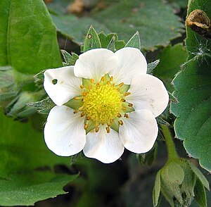
Flower of Garden Strawberry (Fragaria ×ananassa)
Image via Wikipedia

The search for chemical mediators in plant root rhizosphere interactions with symbiotic and pathogenic organisms found in the soil continues to generate interesting research. Martha Hawes group at the University of Arizona reported on the role of sugars, proteins and small molecules found in root cap secretions – a mucilaginous mixture that covers the growing root tip and “converses” with the surrounding matrix of living organisms. The cap is rich in root border cells, which detach from the growing root tip. Curlango-Rivera et al (2010) provides us a bit more detail about which metabolites are biologically active. Neither sugars nor amino acids triggered root growth or border cell production. Transient exposure to biologically active concentration levels of the isoflavonoid pisatin, a phytoalexin, stimulated root border cell production but not root tip growth. I wonder if inhibition of root elongation may “reset” plant growth patterns as root border cells, acting as chemical sense organs, define the nature of the environment?

A second paper used histochemcial methods to profile root metabolites in plants from the Rose family (Hoffman et al., 2010). They found flavan-3-ol molecules in the root tip and border cells. Their findings suggest that the distribution of flavan-3-ols in Fragaria and Malus is under tight developmental control. These molecules are found in plants as catechin and epicatechin derivatives and in long chain (polymeric) form. They influence the taste and medicinal potential of green tea and wine, to name a few well-known plants. Previous researchers summarized their role in chelating toxic cations (metals) in the soil, establishing mycorrhizal interactions and priming plant root defense. This paper suggests a role in the transport of the long distance plant hormone auxin, which would link the chemical cross talk at root border cells with responses that occur in tissue distal to root tips. Hoffman’s research lacked a clear distinction of whether the monomeric or polymeric flavan-3-ol forms where the active species. This has plagued plant research for some time, since the analytical methods for detecting the polymeric forms have been crude and ineffective. All of their samples were from a botanical garden. I wonder if the flavan-3-ol profile would differ compared to native wild grown species?

References:

  1. Curlango-Rivera, G. et al. (2010) Plant Soil 332:267-275
  2. Hoffmann, T. et al. (2011) Plant Biology, 13: no. doi: 10.1111/j.1438-8677.2011.00462.x