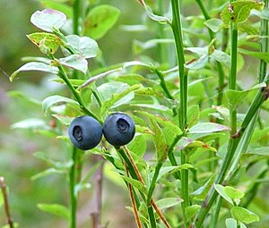
Not Blueberry Pie, but Close

Vaccinium myrtillus
Image via Wikipedia

Bilberry (Vaccinium myrtillus) contains varying levels of phenolic compounds – anthocyanins, chlorogenic acid derivatives, hydroxycinnamic acids, flavonol glycosides, catechins, and proanthocyanidins. Research by Martz et al (2010) elucidated how levels of bilberry leaf phenolics differed along an ecological gradient in boreal forests running north to south in Finland. These regions differ in latitude, altitude, over story cover, levels of continuous light, temperature and associated frost spells.

An analysis of bilberry leaves showed that major phenolic changes in bilberry leaves appeared in the first stages of leaf development. As important, synthesis and accumulation of flavonoids was delayed in the forest compared to the high light sites. Two-fold higher flavonoid levels appeared in leaf tissue growing in high-light intensity sites, higher latitudes, and/or higher altitudes compared to in lower altitudes and low-light intensity sites.

Close and Mcarther (2002) previously theorized that the presence of greater phenolic levels in leaf tissue found in northern regions was a response to colder temperatures, which would limit essential enzyme function, during periods of maximal photo-oxidative stress (Close and Mcarther, 2002).  However, Martz et al (2010) also showed that leaf flavanoid genes were highly expressed in shade, but that the timing of expression appeared to alter the relative metabolite levels in shade compared to sun exposed bilberry leaf.

Mudge et al., (2016), researched phenolic profiles of wild elderberry fruits (Sambucus nigra subsp. canadensis) over two years in eastern US, noting that flavanols (quercitin, isoquercitin, rutin) and chlorogenic acid metabolite concentrations were higher in the southeast, particularly interior. They suggested the variation of phytochemical profiles of the berries were impacted by genetic or environmental factors without understanding on which was more important.

What’s missing from the data picture includes a more complex measurement of ecological influences, such as response to herbivory and rhizosphere fungal associations? This type of whole community data would help to build a more complete picture of  plant response.

This requires sampling, sampling, sampling.

_______________________________________________

  1. Martz, F., Jaakola, L., Julkunen-Tiitto, R. and Stark, S. (2010). Phenolic Composition and Antioxidant Capacity of Bilberry (Vaccinium myrtillus) Leaves in Northern Europe Following Foliar Development and Along Environmental Gradients. J Chem Ecol, published online, 19 August 2010
  2. Close, D.C., and Mcarther, C. (2002). Rethinking the role of many plant phenolics—protection from photodamage not herbivores? Oikos. 99:166–172
  3. Mudge, E., Applequist, W. L., Finley, J., Lister, P., Townesmith, A. K., Walker, K. M., & Brown, P. N. (2016). Variation of Select Flavonols and Chlorogenic Acid Content of Elderberry Collected Throughout the Eastern United States. Journal of food composition and analysis : an official publication of the United Nations University, International Network of Food Data Systems, 47, 52–59.

Rhizosphere Influence on Plant Medicine

Einjähriger Beifuß (Artemisia annua)
Artemisia annua
Wikipedia

Mycorrhization leads to nutrient and information flow, often in both directions. The plant root supplies sugars to the fungus, while the fungus induces Jasmonic Acid biosynthetic enzymes in the plant, leading to an increase in jasmonate ­ levels that enhance the accumulation of soluble sugars in plant root and the production  of plant root defense compounds.

From a research article,  the presence of mycorrhizal fungus, Glomus mosseae and nitrogen fixing Bacillus subtilis on the roots influenced the levels of plant biomass growth, and the yield of an important medicinally active phytochemical, artemisinin, from Artemisia annua L and used as an anti-malarial treatment.

Gabriele et al. (2016) investigated the effect of mycorrhizal soil inoculation of various Sangiovese wine grapes and found the presence of the fungus increased levels of 14 polyphenols compared to un-inoculated plants. Here the presence of symbiotic relations in the soil altered the phytochemical makeup of fruit.

So how are the plant roots attracting mycorrhizal symbionts? Plant produced flavanoid compounds accumulate at root tips/cap and make up a large portion of root exudate (the portion of the root sap excreted to the external environment). These phytochemicals are easily modified and their biosynthesis is triggered  by transcription factors, which suggests a role as elicited signal compounds – compounds that are made specifically in response to conversation from rhizosphere fungi and bacteria. Interestingly, their presence in the rhizosphere soil triggers mycorrhizal fungi to explore their surroundings (Hassan and Mathesius, 2012), perhaps increasing the likely hood of contact with plant roots.

Given the high price of American wild grown ginseng, the ecological influence on ginsenoside formation, and ultimately, the therapeutic value, points to optimizing the rhizosphere cross talk by way of forest farming.

The highest ginsenoside content occurs (from highest to lowest) in the root hairs > lateral roots > cortex > interior taproot (Li and Wardle, 2002), exactly where we should expect a chemical conversation to occur.

Within this class of compounds we designate as ginsenosides, two molecular forms are dominant, protopanaxadiols and protopanaxatriols. Data from two different papers (Zhu et al., 2004: Wang et al., 2010) compared levels of diols and triols in different species and sources of ginseng. American ginseng (Panax quinquefolia) had higher levels of the triols (especially Rg1) compare to Chinese ginseng (P. ginseng), which had higher levels of diols (especially Rb1  Rd).

Structures-of-ginsenosides-from-Panax-ginseng-Glc-glucose-Rha-rhamnose-Araf
Li, H, Lee, JH, and Ha, JM. (2008) Effective Purification of Ginsenosides from Cultured Wild Ginseng Roots, Red Ginseng, and White Ginseng with Macroporous Resins. Journal of Microbiology and Biotechnology. 18(11):1789-91. DOI: 10.4014/jmb.0800.192

Comparing wild grown versus cultivated plants within each species, a similar pattern emerged, with wild plants showing a higher concentration of triols (especially Rg1  Re), while cultivated plants had higher concentration of diols (especially RbRb2).

James, et al. (2013) investigated levels of diols and triols in wild sourced P. quinquefolia leaf and root  in a North Carolina collection, finding that there was no relationship between age and ginsenoside content. However total ginsensosides were higher in the leaf, as was Rb2 and Rd (diols), In the root tissue, Rb1(diol) and Rg1 (triol) was found to be higher.

This has implications for how we “farm” medicine and speaks to a long held tenet; complex interactions in native ecologies, including the soil,  produce medicinal plant crops that are more biologically active. Farm versus wild grown ginseng is only one example. What’s been your experience as a imbiber, herbalist, researcher, plant grower or manufacturer?


Do Plant Roots Talk to Leaves?

Arabidopsis thaliana
Arabidopsis thaliana (Wikipedia)

Surrounded by material excreted (exudate) by their own root border cells, the growing root tips (apical region) of plants move through soil regions where important biological interactions occur with a community of soil microbes. This exudate not only helps define the soil microbiome (microbial community), but also changes the physical and chemical characteristics of rhizosphere soil.

Root-tip-tag
Root tip (100×) 1. Meristem 2. Columellae 3. Lateral part of the tip 4. Dead cells 5. Elongation zone (Photo: SuperManu – Clematis)

Hiltpold et al (2011) provided evidence of  systemic, volatile signals in maize roots in response to herbivore attack. From 2013 research on Arabidopsis suggests that soil microbes can alter plant leaf chemistry to inhibit insect feeding. They posited a role for microbial-derived volatile organic compounds acting as a deterrence signal, and noted the presence of Actinobacteria, Firmicutes and Proteobacteria in soil and within Arabidopsis root tissue.In a 2013 Tansley Review, Turnbull and Lopez-Cobello noted that despite localized cellular communication found in the root apical meristem, communication via vascular transport to the rest of the plant did not seem to occur. That left me wondering how plant roots communicated changes throughout the entire plant (systemic).

Those microbes are often associated with “soil odors”. On a sensorial level,  “smelling” the earth may help us appreciate the complex, unseen communication happening under foot.